


Understanding radiology
- A radiology department is where your imaging scan test will take place.
- A radiographer will perform your imaging scan. They have technical knowledge of the equipment.
- A radiologist will look at your results and tell you or your doctor what they mean.
Chest X-ray
Chest X-rays are normally done in a hospital radiology department. It will only take a few minutes.
X-rays are a type of radiation. The radiation from a chest X-ray is very small and the risk from the radiation is very low.
What is a chest X-ray used for?
A chest X-ray creates an image of the inside of your chest. It shows your lungs, ribs, heart, and diaphragm. You may need a chest X-ray if you have breathlessness, chest pain, or you are coughing up blood. A chest X-ray can also see if you have a lung infection, like pneumonia.
How can I prepare for a chest X-ray?
Your appointment letter should tell you if there’s anything you need to do to prepare for your chest X-ray. You usually don’t need to do anything to prepare for a chest X-ray.
Try to avoid wearing anything metal to your appointment, such as:
- jewellery
- zips
- bras with metal underwire
- belts.
If you’re pregnant, or think you could be, it’s important to tell the radiographer, so that they can reduce radiation exposure to your unborn baby.
What happens during a chest X-ray?
Before your chest X-ray, you might need to take off any clothes on your top half, including shirts, vests, and bras. You may be asked to wear a hospital gown too.
When you’re ready, the radiographer will leave the room or stand behind a screen to control the X-ray machine. The test will only take a few minutes.
You’ll usually be standing up, though chest X-rays can be taken sitting or lying down. The X-ray machine points at your chest to take pictures. You need to hold your breath for a few seconds while the picture is taken. This makes sure that the image isn’t blurred.
CT scan
Computerised tomography scans (CT) are normally done in a hospital radiology department. They use X-rays to build a detailed picture of the inside of your body, including your lungs, blood vessels and other organs.
What is a CT scan used for?
CT scans are used to understand what’s going on inside your lungs. They can see if your lungs are healthy and can help to diagnose lung conditions. A CT scan might also be used to decide what type of treatment you need.
Sometimes a CT scan might be used to monitor conditions or understand if your treatment is working - for example, in people with lung cancer.
You might also have a CT scan of your nose and sinuses to check for inflammation.
How can I prepare for a CT scan?
You should get a letter telling you how to prepare for your CT scan. Read this carefully to see if there are any instructions. You may be told to:
- not eat two hours before your appointment
- not wear any metal like jewellery, belts, underwired bras, or zips.
Tell hospital staff if you are allergic to iodine, as the contrast dye injected as part of some scans may have iodine in.
If you are pregnant, you should not have a CT scan, as this could harm your unborn baby.
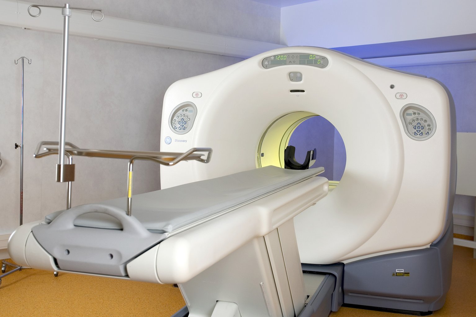
What happens during a CT scan?
During a CT scan, you lie on a cushioned table. The table slides into a circular scanner. The scan will take around 10-20 minutes. A radiographer will stay in the next room and control the scanner.
Let the radiographer know if you feel worried. They’ll be able to answer any questions you have. You will go home the same day.
You may be injected with a special dye, called contrast. This will help to improve the quality of the CT scan image.
The dye is normally harmless and will leave your body through your pee. Some people might have an allergic reaction to the dye. Don’t worry though, your radiologist is trained to deal with allergic reactions.
PET scan
Positron emission tomography scans (PET) are normally done in a hospital radiology department.
PET scans measure cell activity in different parts of your body. The scan creates detailed 3-D images of the inside of your body. Sometimes a PET scan is used with a CT scan to create a more detailed image. This is called a PET-CT scan.
What is a PET scan used for?
A PET scan will see if there is inflammation, infection, or scarring in your lungs. It is also used to check for lung cancer.
If you have been diagnosed with lung cancer, a PET scan can see if cancer treatments are working. Regular PET scans are important to make sure cancer has not spread to another part of the body. PET scans are important for planning the best treatment too.
How can I prepare for a PET scan?
You’ll get a letter from the hospital telling you how to prepare for your PET scan. You'll usually be told not to eat anything for six hours before, but you can drink water. You should also avoid intense exercise for 24 hours before your appointment.
You need to arrive on time for your PET scan. This is because the radioactive liquid used in the scan is prepared before the appointment, and only works for a short amount of time. If you’re late, your scan may be cancelled, and you’ll have to wait for another appointment.
What happens during a PET scan?
Before the scan, you’ll be injected with a liquid, called a radiotracer. This is slightly radioactive and can be seen by the scanner. The radiotracer is safe and will leave your body within a few hours.
You will lie on a cushioned table with a large, circular scanner at one end. The cushioned table will slide into the PET scanner. It’s important that you stay as still as you can while you are in the scanner, to stop the image from blurring.
PET scans are painless and take around 30 to 60 minutes.
After your PET scan
You’ll be asked to avoid pregnant people, babies, and children for a few hours after your PET scan. This is while the radiation leaves your body. Drinking plenty of water will help the radiotracer to leave your body. If you have children, it’s important to organise childcare to cover this time.
VQ scan
A VQ scan is also called a ventilation-perfusion scan or a lung perfusion scan. It looks at the flow of air and blood in your lungs. They’re normally done in a hospital radiology department.
What is a VQ scan used for?
The most common use for a VQ scan is to see if there are blood clots (pulmonary embolism) in the lungs. They can help to find out which part of your lungs need treatment.
VQ scans are also used to help plan operations to remove lung cancer. If you have emphysema, a VQ scan can help find out if you can have lung volume reduction surgery.
The VQ scan is sometimes done with a CT scan to get a more detailed picture.
How can I prepare for a VQ scan?
You should get a letter telling you how to prepare for your V/Q scan. Read this carefully to see if there are any instructions.
If you’re pregnant, you might be told to have the VQ scan after your baby is born. This is to protect your unborn baby from the radiation.
What happens during a VQ scan?
A VQ scan includes two tests:
- Ventilation - you will breathe in a slightly radioactive gas called krypton for about 10 minutes. This does not smell and you can’t taste it either.
- Lung perfusion - you will have an injection of slightly radioactive material. Sometimes you will only need this part of the scan.
The scan takes around one hour. The amount of radiation in the gas and injection is not dangerous.
Your results
You won’t usually get the results of any imaging scans straight away - unless you are unwell in hospital. A specialist radiologist will review your scans. Your doctor will contact you if the results of your scan are abnormal.

Get support
Call our Helpline for support with your condition. Get advice on your medicines, symptoms or travelling with a lung condition, or just call us to say hello.